陰部神經

 Clash Royale CLAN TAG#URR8PPP
body.skin-minerva .mw-parser-output table.infobox captiontext-align:center
| 陰部神經 | |
|---|---|
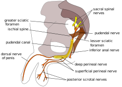 男性骨盆的陰部神經路徑圖。 | |
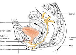 女性骨盆的陰部神經路徑圖,陰部神經來自第2、3、4對骶部脊神經(S2、S3、S4),由子宮及肛門之間延伸進入小陰唇、大陰唇及陰蒂 | |
| 细节 | |
| 拉丁语 | Nervus pudendi |
| 來源 | 薦神經(S2 ~ S4) |
| 走向 | 下直腸神經 會陰神經 陰莖背神經 陰蒂背神經 |
| 识别标示 | |
| Gray's | p.967 |
| Dorlands | _05/12566568 |
| TA | A14.2.07.037 |
| FMA | FMA:19037 |
解剖学术语 | |
陰部神經(英语:pudendal nerve)為會陰部的主要神經[1]:274。在感覺方面,該神經負責傳送男性及女性外陰部、肛門周圍,以及會陰的感覺信息;運動信息方面,則支配了男性及女性的尿道括約肌,和外肛門括約肌。一旦損傷則可能導致排糞失禁,這類損傷常見於分娩後遺症,麻醉也可能造成類似的症狀。
陰部神經會經過閉孔內肌上的陰部管。1836年,愛爾蘭解剖學家本傑明·阿爾科克首次描述陰部管,故該管又稱阿爾科克氏管(英语:Alcock's canal)。
目录
1 解剖
1.1 神經核
1.2 變異
2 功能
3 臨床意義
3.1 麻醉
3.2 損傷
3.3 影像
3.3.1 神經傳導潛伏期試驗
4 歷史
5 其他圖片
6 參見
7 參考文獻
8 外部連結
解剖

圖中可看到坐骨大孔及坐骨小孔,兩者以骶棘韧带隔開。陰部神經由坐骨大孔離開骨盆,越過韧带,再經由坐骨小孔進入骨盆
陰部神經共有一對,分別在身體的左右側。每一邊的神經都是由三根神經在骶棘韧带及尾骨肌的上邊界上方合成一根神經[2]。這三根神經中,中間的及下方的神經合成下方的神经索,因此變成二條神经索,再在骶棘韧带附近聯合成一條陰部神經[3]。這三條神經源自第2、3、4對骶部脊神經的腹支,主要是來自第4對骶部脊神經[2][4]:215[5]:157。
陰部神經通過梨狀肌及尾骨之間的部位,從坐骨大孔的下方離開骨盆[2]。陰部神經通過骶棘韧带的外側,從坐骨小孔再進入骨盆,之後會伴隨著陰部內動脈及陰部內靜脈,沿著坐骨直腸窩的側壁往上往前,和內動脈和內靜脈包覆在閉孔肌筋膜的鞘中,稱為阴部管[6]:8。
陰部神經在陰部管內會分支,先分支為內直腸神經,之後是會陰神經,最後是男性的陰莖背神經或是女性的陰蒂的背神經[6]:34。
神經核
此神經是骶叢的主要分支之一[7]:950,神經纖維起源自骶骨段脊髓的歐氏神經核[3]。
變異
陰部神經源自的神經位置也可能會變化,例如有些人的陰部神經可能是源自坐骨神經[8]。,因此坐骨神經的損傷也會影響陰部神經。有時第1節骶神經的後支也可能發展成為陰部神經,甚至是更少見的第5節骶神經(S5)[3]。
功能
陰部神經同時具有運動及感覺兩種功能,在自律神經系統方面僅有交感神經的神經纖維,沒有副交感神經的纖維[9]:1738。
男性的陰部神經會分出陰莖背神經支配陰莖的感覺,女性則會分出陰蒂背神經支配陰蒂感覺[10]:422。男性的陰囊後側由陰囊後神經支配其感覺,女性對應的陰唇後神經會支配陰唇的感覺。這些部位有許多神經傳導其感覺,陰部神經即為其中之一[11]。陰部神經也傳導肛管部位的感覺[6]:8。陰部神經會傳導陰莖及陰蒂的感覺,因此也是在陰莖勃起及陰蒂勃起過程中的传入神经[12]:147。陰部神經也負責射精相關的功能[13]。
陰部神經的分支也支配會陰及骨盆底的肌肉,包含球海绵体肌、坐骨海绵体肌[11],以及提肛肌(包括腸骨尾骨肌、恥骨尾骨肌、耻骨直肠肌、女性的恥骨陰道肌或是男性的前列腺提肌)[10]:422[14]、外肛門括約肌(透過下肛門分支)[6]:7、以及男性尿道外括約肌或女性尿道外括約肌[10]:424–425。
陰部神經還透過乙醯膽鹼釋放控制尿道外括約肌的肌張力。當乙醯膽鹼的釋放量增加,尿道外括約肌內的骨骼肌纖維會收縮,使尿液留在膀胱,反之則能促進排尿[15]。
臨床意義
麻醉
陰部麻醉也稱為阴部神经阻滞,或鞍神经阻滞(saddle nerve block),是產科使用的局部麻醉,可在分娩時麻醉陰部[16]。此麻醉方式會在陰道內壁注射利多卡因,目的是要影響陰部神經[17]。
損傷
陰部神經可能會被壓縮或是伸展,造成暫時或是永久的神經病變。若陰部神經拉伸了原來長度的12%,可能會造成不可逆的神經受損[6]:655。若盆腔底急性過度拉伸(例如滯產或是難產)或慢性過度拉伸(因便秘造成排便時的慢性拉伸),可能會讓陰部神經出現拉伸造成的神經病變[6]。陰部神經卡壓也稱為阿爾科克氏管症候群(Alcock canal syndrome),是非常少見的疾病,多半發生在職業的自行車選手身上[18]。像糖尿病及多发性硬化症等系統性疾病也可能透過脫髓鞘病或是其他機制使陰部神經受損[6]:37。骨盆腔的腫瘤(最著名的是大型的骶尾部畸胎瘤)或是去除腫瘤的手術都可能造成神經永久的受損[19]。
若單側的陰部神經病變可能會造成糞便失禁,但也有例外[6]:34。
影像
用一般的斷層掃描或是核磁共振成像,很難對陰部神經顯像。不過透過斷層掃描的引導,可以將針插到鄰近陰部神經血管束的部位。坐骨棘在斷層掃描時很容易識別,因此會插在此一部位。脊椎針會通過臀肌前進,在坐骨棘上前進幾個毫米。之後會注射X光的顯影劑,讓阴部管內的神經更加清楚,也可以確認針插入的位置是否正確。然後會注射可的松到神經中,進行局部麻醉進行確認,也治療外陰部的慢性疼痛(女性稱為外阴疼痛)、骨盆疼痛和肛門直腸疼痛等[20][21]。
神經傳導潛伏期試驗
陰部神經的延遲時間可以量化,具體的定義是從在感覺神經給電刺激的時間起,到運動神經有訊號使陰部肌肉收縮的時間,時間太長代表神經受損[22]:46。測試時會有兩個固定在手指端的刺激電極及兩個量測電極(St Mark電極)[22]:46。
歷史
陰部神經的拉丁文為「Nervus pudendus」。「Nervus」一詞指的是神經;「Pudenda」一詞來自拉丁文,意即外生殖器官,乃源自「pudendum」這個字,意思是「帶來羞恥的部份」[23]。陰部管也稱為阿尔科克氏管(Alcock's canal),得名自1836年首次紀錄此一部位的愛爾蘭解剖學家班傑明·阿爾科克。阿爾科克在羅伯特·本特利·托德的《生理暨解剖學百科全書》(The Cyclopædia of Anatomy and Physiology) 一書中,於描述髂動脈群的章節內首次提及陰部神經及陰部管[24]。
其他圖片

男性骨盆,陰部神經位於圖中右方。

陰部神經支配構造模式圖。

男性骨盆的陰部神經路徑圖。


參見
本條目使用了部分解剖術語,概述請參閱這裡。
- 神經性膀胱功能異常
參考文獻
^ AMR Agur, AF Dalley, JCB Grant. Grant's atlas of anatomy 13th. Philadelphia: Wolters Kluwer Health/Lippincott Williams & Wilkins. 2013. ISBN 978-1-60831-756-1.
^ 2.02.12.2 Standring S (editor in chief). Gray's Anatomy: The Anatomical Basis of Clinical Practice 39th. Elsevier. 2004. ISBN 978-0-443-06676-4.
^ 3.03.13.2 Shafik, A; el-Sherif, M; Youssef, A; Olfat, ES. Surgical anatomy of the pudendal nerve and its clinical implications. Clinical Anatomy. 1995, 8 (2): 110–5. PMID 7712320. doi:10.1002/ca.980080205.
^ Moore, Keith L. Moore, Anne M.R. Agur ; in collaboration with and with content provided by Arthur F. Dalley II ; with the expertise of medical illustrator Valerie Oxorn and the developmental assistance of Marion E. Essential clinical anatomy 3rd. Baltimore, MD: Lippincott Williams & Wilkins. 2007. ISBN 978-0-7817-6274-8.
^ Russell RM. Examination of peripheral nerve injuries an anatomical approach. Stuttgart: Thieme. 2006. ISBN 978-3-13-143071-7.
^ 6.06.16.26.36.46.56.66.7 Wolff BG et al. (编). The ASCRS textbook of colon and rectal surgery. New York: Springer. 2007. ISBN 0-387-24846-3.
^ TL King; MC Brucker; JM Kriebs; JO Fahey. Varney's midwifery Fifth. Jones & Bartlett Publishers. 2013. ISBN 978-1-284-02542-2.
^ Nayak, Soubhagya R.; Madhan Kumar, S.J.; Krishnamurthy, Ashwin; Latha Prabhu, V.; D'costa, Sujatha; Jetti, Raghu. Unusual origin of dorsal nerve of penis and abnormal formation of pudendal nerve—Clinical significance. Annals of Anatomy - Anatomischer Anzeiger. November 2006, 188 (6): 565–566. doi:10.1016/j.aanat.2006.06.011.
^ Neill, editor-in-chief, Jimmy D. Knobil and Neill's physiology of reproduction 3rd. Amsterdam: Elsevier. 2006. ISBN 0-12-515400-3.
^ 10.010.110.2 Drake, Richard L.; Vogl, Wayne; Tibbitts, Adam W.M. Mitchell; illustrations by Richard; Richardson, Paul. Gray's anatomy for students. Philadelphia: Elsevier/Churchill Livingstone. 2005. ISBN 978-0-8089-2306-0.
^ 11.011.1 Ort, Bruce Ian Bogart, Victoria. Elsevier's integrated anatomy and embryology. Philadelphia, Pa.: Elsevier Saunders. 2007. ISBN 978-1-4160-3165-9.
^ Babayan, Mike B. Siroky, Robert D. Oates, Richard K. Handbook of urology diagnosis and therapy 3rd. Philadelphia, PA: Lippincott Williams & Wilkins. 2004. ISBN 978-0-7817-4221-4.
^ Penson, David F. Male Sexual Function: A Guide to Clinical Management. Annals of Internal Medicine. 2002.
^ Guaderrama, Noelani M.; Liu, Jianmin; Nager, Charles W.; Pretorius, Dolores H.; Sheean, Geoff; Kassab, Ghada; Mittal, Ravinder K. Evidence for the Innervation of Pelvic Floor Muscles by the Pudendal Nerve. Obstetrics & Gynecology. October 2005, 106 (4): 774–781. doi:10.1097/01.AOG.0000175165.46481.a8.
^ Fowler, CJ; Griffiths, D; de Groat, WC. The neural control of micturition. Nat. Rev. Neurosci. June 2008, 9: 453–66. PMC 2897743. PMID 18490916. doi:10.1038/nrn2401.
^ Lynna Y. Littleton; Joan Engebretson. Maternal, Neonatal, and Women's Health Nursing, Volume 1. Cengage Learning. 2002: 727.
^ Satpathy, Hemant K.; 等. Isaacs, Christine; 等, 编. Transvaginal Pudendal Nerve Block. WebMD LLC. [2015-07-19].
^ Mellion MB. Common cycling injuries. Management and prevention. Sports Med. January 1991, 11 (1): 52–70. PMID 2011683. doi:10.2165/00007256-199111010-00004.
^ Lim, Jit F.; Tjandra, Joe J.; Hiscock, Richard; Chao, Michael W. T.; Gibbs, Peter. Preoperative Chemoradiation for Rectal Cancer Causes Prolonged Pudendal Nerve Terminal Motor Latency. Diseases of the Colon & Rectum: 12–19. doi:10.1007/s10350-005-0221-7.
^ Calvillo O, Skaribas IM, Rockett C.; Skaribas; Rockett. Computed tomography-guided pudendal nerve block. A new diagnostic approach to long-term anoperineal pain: a report of two cases. Reg Anesth Pain Med. 2000, 25 (4): 420–3. PMID 10925942. doi:10.1053/rapm.2000.7620.
^ Hough DM, Wittenberg KH, Pawlina W, Maus TP, King BF, Vrtiska TJ, Farrell MA, Antolak SJ Jr.; Wittenberg; Pawlina; Maus; King; Vrtiska; Farrell; Antolak Jr. Chronic perineal pain caused by pudendal nerve entrapment: anatomy and CT-guided perineural injection technique. Am J Roentgenol. 2003, 181 (2): 561–7. PMID 12876048. doi:10.2214/ajr.181.2.1810561.
^ 22.022.1 G.A. Santoro, A.P. Wieczorek, C.I. Bartram (editors). Pelvic floor disorders imaging and multidisciplinary approach to management. Dordrecht: Springer. 2010. ISBN 978-88-470-1542-5.
^ Harper, Douglas. Pudendum. Online Etymology Dictionary. [2014-02-28].
^ Oelhafen, Kim; Shayota, Brian J.; Muhleman, Mitchel; Klaassen, Zachary; Tubbs, R. Shane; Loukas, Marios. Benjamin Alcock (1801-?) and his canal. Clinical Anatomy. 2013-09, 26 (6): 662–666. PMID 22488487. doi:10.1002/ca.22080.
外部連結
维基共享资源中相关的多媒体资源:陰部神經 |
Anatomy figure: 41:04-11 at Human Anatomy Online, SUNY Downstate Medical Center - 女性會陰下視觀,以及內陰動脈的分支
figures/chapter_32/32-2.HTM — Basic Human Anatomy at Dartmouth Medical School
figures/chapter_32/32-3.HTM — Basic Human Anatomy at Dartmouth Medical School
橫截面圖像:pelvis/pelvis-female-17 - 維也納醫科大學生物塑化實驗室提供- www.nervemed.com 上的診斷與治療方法
- www.pudendal.com
- 陰部神經壓迫症候群 - chronicprostatitis.com
- 陰部神經阻滯的電腦斷層影像
| ||||||||||||||||||||||||||||||||||||||||||||||||||||||||||||||||||||||||||||||||||||||||||
